Développement professionnel
Apprendre. Partager. Progresser
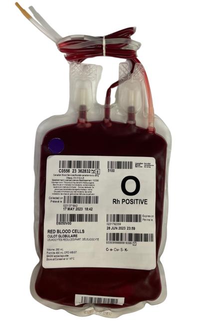
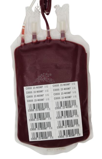
Culots globulaires: Hémolyse
Légende de couleurs
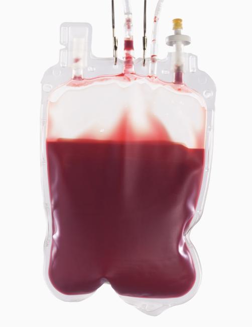
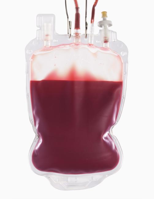
Exemple(s) d’aspect typique
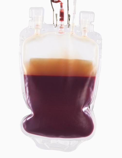
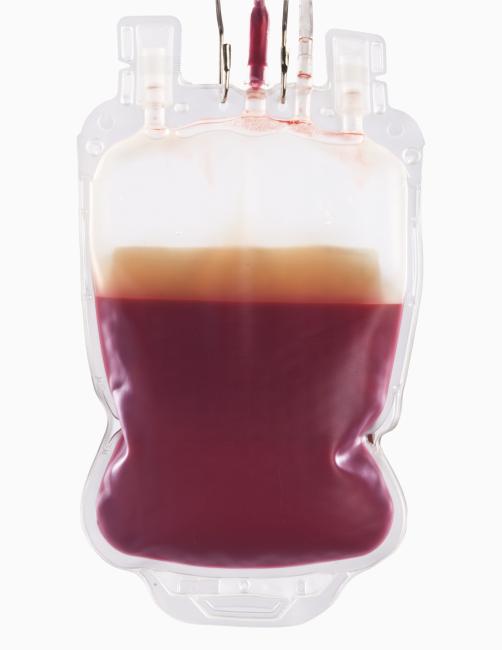
Exemple(s) de changement d’aspect
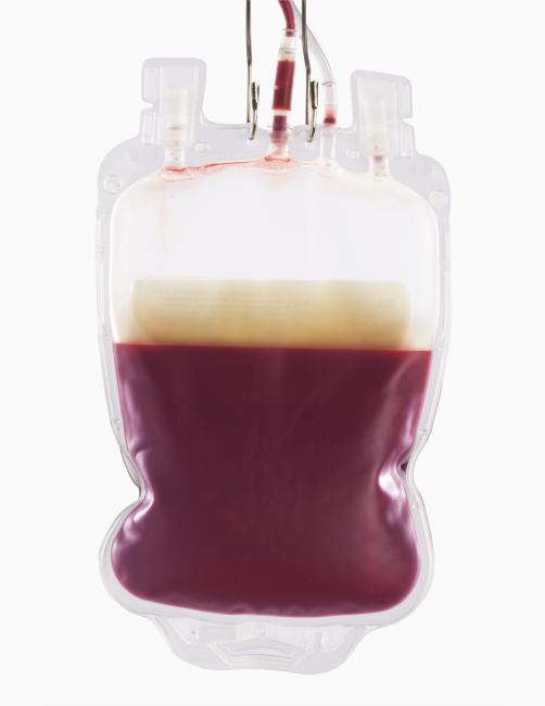
L’hémolyse se produit lorsqu’il y a rupture des globules rouges, puis libération du contenu intracellulaire dans le liquide surnageant. La libération de l’hémoglobine (protéine pigmentée des globules rouges) a une influence importante sur l’aspect des culots globulaires et de leurs segments. L’hémolyse des culots globulaires entreposés est un processus normal qui s’accentue avec la durée de l’entreposage. Un certain niveau d’hémolyse est acceptable et prévisible.
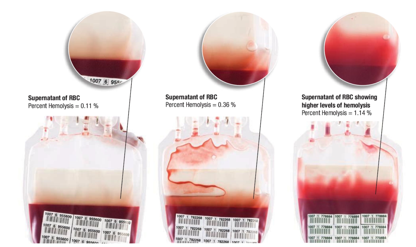
L’Association canadienne de normalisation (CSA) fixe la limite inacceptable d’hémolyse à ≥ 0,8 % de la masse de globules rouges dans 90 % des unités testées à la date de péremption. L’inspection visuelle de la couleur du liquide surnageant reste toutefois le seul moyen non destructif d’estimer l’hémolyse dans un culot globulaire. C’est une méthode très subjective, et il est difficile de définir une couleur limite pour l’acceptabilité d’une unité. La couleur du liquide surnageant permet d’estimer la concentration en hémoglobine. Toutefois, le pourcentage d’hémolyse d’un culot globulaire est également influencé par d’autres facteurs que la concentration en hémoglobine comme l’hématocrite ou l’hémoglobine totale. Par exemple, une augmentation de l’hématocrite ou de l’hémoglobine totale entraînera un plus faible pourcentage d’hémolyse lorsque l’hémoglobine du liquide surnageant est maintenue à un niveau constant. La concentration en hémoglobine du liquide surnageant est donc l’un des seuls facteurs pouvant être pris en compte pour déterminer le pourcentage d’hémolyse d’un culot globulaire.
Image
![Percent hemolysis equals [(100 - hematocrit) x supernatant hemoglobin (g/L)] divided by total hemoglobin (g/L)](/sites/default/files/2024-01/Fig1_Hemolysis%20calculation.jpg)
La présence de globules rouges dans le liquide surnageant peut à tort être prise pour de l’hémolyse. Si l’on soupçonne une hémolyse, il peut être utile, avant toute vérification, de laisser l’unité se stabiliser pendant au moins 24 heures pour limiter la contamination du liquide surnageant par les globules rouges.
Plusieurs causes expliquent l’hémolyse. Voici quelques exemples liés au processus de fabrication et à la manipulation des culots globulaires :
- Exposition à des températures extrêmes
- Transformation du produit
- Contamination bactérienne
- Manipulation inadéquate
- Solutions incompatibles
Aspect
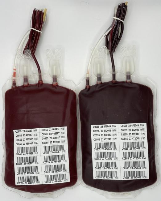
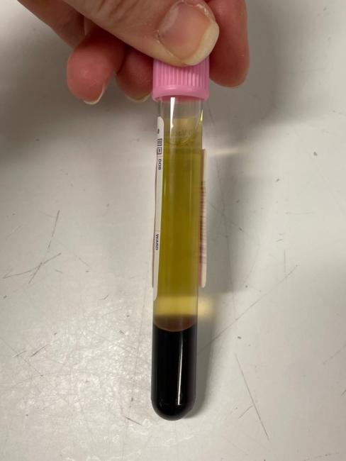
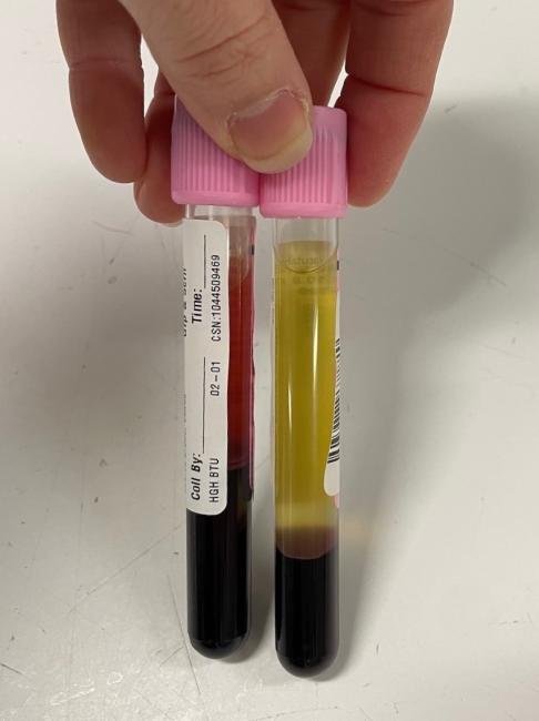
- La diminution du nombre de globules rouges intacts se traduit par un hématocrite plus faible et une couleur rouge plus vive.
- L’hémolyse causée par la contamination bactérienne peut entraîner une altération de la couleur allant du mauve foncé au noir (voir la section sur la contamination bactérienne).
- L’hémoglobine libre donne au liquide surnageant une teinte allant du rose pâle au rouge foncé presque mauve, selon l’étendue de l’hémolyse (une hémolyse plus importante donnera une couleur plus foncée).
- Note : Les segments ne reflètent pas avec exactitude l’hémolyse du culot et ne doivent pas être utilisés pour estimer l’hémolyse.